Shedding light on the nanoworld
For years, scientists thought that optical microscopes - which work using light from the visible part of the spectrum - had reached the limit of their resolution. Because light diffracts around anything smaller than half its own wavelength, it was assumed that it would be impossible to clearly view anything smaller than this size, such as the contents of our cells.
The development of fluorescence microscopy changed all that. The technique involves marking molecules with fluorescent chemicals – known as fluorophores - which glow when light is applied.
Professor Hell's innovation was to illuminate the sample by training two separate lasers on it - one to make the fluorescent molecules glow and another to switch off the glow in all but a nanometre-sized area. By repeating this process across the whole sample, he could build up a comprehensive image nanometre-by-nanometre.
Cancer detection
One promising application of the technique is the early detection of cancer. Professor Jerker Widengren, from the KTH Royal Institute of Technology in Sweden has been able to identify unique features on the surface of cancer cells and devise fluorescently marked molecules to bind with them.
‘Our first objective was to identify breast and prostate tumours,’ said Prof. Widengren, who coordinated the EU-funded FLUODIAMON project, which ran from 2008 to 2012. During this project his team was able to show that the fluorescence-based approach requires minimal amounts of human tissue for an accurate diagnosis, reducing pain and the risk of infection for patients.
“‘If this discovery has taught us anything, it is that one should keep an open mind.’
They are now investigating whether the technique can be used to detect the early signs of cancer from a blood sample. ‘We think that tumours may build their own blood vessels by stimulating nearby platelets,’ said Prof. Widengren. ‘Fluorescence microscopy is the first technique that can resolve the protein distribution in these cells to show us what is going on.’
He says the potential for the technology is vast. ‘The wavelength, intensity and polarisation of the light emitted by fluorophores can also provide details on the chemical microenvironment of the molecules.’
Into the cell
Thanks to its super-high resolution, fluorescence microscopy also enables us to view sub-cellular structures such as DNA. Professor Johan Hofkens at the University of Leuven in Belgium leads the FLUOROCODE project, which is using a variation of the technique to sort through DNA and understand more about how mistakes in genetic read-out could be linked to cancer.
Although genetic sequencing is getting faster and cheaper by the day, Prof. Hofkens says that assembling DNA readouts remains a painstaking task. The FLUOROCODE project, which is funded by the European Research Council (ERC), is using fluorescence microscopy to map out entire genomes before they are broken down for sequencing.
Researchers on the project have devised a highly efficient method to label specific DNA sequences in our genome with fluorescent chemicals. These fluorophores can also reveal precious information about their surroundings.
‘Food, stress and even pollution can cause molecules to bind in irregular ways with our DNA,’ said Prof. Hofkens. ‘This can dangerously affect the way in which the cell reads out its genetic code.’ He is now working with a biotech company to commercialise a diagnostics tool.
However, Prof. Hofkens says the greatest legacy of the development of fluorescence microscopy may lie not in its technological breakthroughs but in changing the mind-set of the research community. ‘If this discovery has taught us anything, it is that one should keep an open mind,’ he said. ‘I feel privileged to work with a community and funding agencies that allow young scientists to push for new ideas.’
- 2014The Nobel Prize and beyond
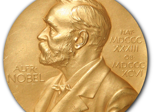
The Nobel Prize in Chemistry is awarded to Professor Stefan Hell, Dr Eric Betzig and Professor William Moerner for taking optical microscopy into the nanodimension. Scientists can now observe the interaction of individual molecules inside cells, see how molecules create synapses between nerve cells in the brain, track proteins involved in Parkinson's, Alzheimer's and Huntington's diseases as they aggregate, follow individual proteins in fertilized eggs as these divide into embryos, and much more. Further advances could lead to 3D films of living cells.
 Dr Eric Betzig and Professor William Moerner shared the Nobel Prize with Professor Stefan Hell for separate work in devising another method of circumventing Abbe’s limit in fluorescence microscopy.


